At a long COVID clinic, here’s how doctors are trying to help one woman who is struggling

Belinda Hankins first grappled with COVID-19 in the spring of 2020. She had a fever, chills and trouble breathing, but the real clincher was her loss of smell. Hankins remembers opening a canister of Tony Chachere’s creole seasoning, lowering her nose to take a whiff, and not smelling a thing. “That stuff usually clears the kitchen,” she says.

Her second infection, two years later, was worse. After 12 long weeks of endless fatigue and aching joints, her doctor suggested she seek treatment for long COVID. The lingering, sometimes full-body condition can plague people for months or years after a COVID-19 infection (SN Online: 7/29/22).

In late August, I joined Hankins, age 64, in a small exam room for her first in-person consultation at the Johns Hopkins Post-Acute COVID-19 Team clinic. Wearing a navy dress and a blue surgical mask, Hankins is sitting in a chair across from physician Alba Azola. As they discuss Hankins’ symptoms, doctor and patient face each other, Azola occasionally swiveling her stool to tap notes into a computer.

Hankins’ symptoms are extensive. Brain fog, fatigue and pain top the list. She’s depressed. Sleep doesn’t feel restful. She has trouble focusing, is often light-headed and regularly loses her balance. Even walking to the clinic from the parking lot left her winded and in pain. “I’m extremely exhausted,” she says. “I have not felt good in a long time.” Hankins, pauses, wiping away a tear. “I wasn’t like this before.”
Hankins, a retired digital media consultant, used to be an avid skier and a cyclist. She loved to travel and dance and was planning to learn how to play golf. She’s not sure what the future holds, though she tells me she still has faith she can be active again.

Treating people with long COVID can be complicated – especially for Hankins and those who have other medical conditions. She has pulmonary hypertension, fibromyalgia and the connective tissue disease scleroderma. It’s tricky to tease out which symptoms come from the viral infection. Azola’s approach is to listen, ask questions and listen some more. Then, she’ll zero in on a patient’s most pressing concerns. Her goal: manage their symptoms. “How can we make their quality of life better?” she asks.
System overload
On the afternoon of Hankins’ visit, it’s a warm summer day in Baltimore, blue skies laden with fleecy clouds. Inside the labyrinthine halls of Johns Hopkins Bayview Medical Center, the vibe is not quite as sunny: bright lights, shiny floors, people in line and people in scrubs. Everyone I see is masked.

Azola meets me in the waiting area, walking briskly and wearing bright red glasses. Before the pandemic, Azola, a rehabilitation physician, treated patients recovering from strokes, spinal cord injuries and other disorders. Most mornings, she still works with these patients. But for the past two years, her afternoons have been booked with people laid low by COVID-19.

She’s squeezed me in to talk about the Johns Hopkins PACT clinic, which opened in April 2020, around the time when the world hit one million confirmed cases. “To be honest, we didn’t know what to expect,” Azola says. Back then, most of the clinic’s patients were recovering from COVID-19 after a stay in the hospital’s intensive care unit. Now, at least half of their patients never got sick enough with COVID-19 to be hospitalized – yet still had symptoms they couldn’t shake. In a single week, Azola and her colleagues may get 30 referrals. “It’s constant,” she says, “more than we can provide service to.”

As those referrals pile up, patient wait times can stretch. The PACT clinic expanded last summer, and now has more than a dozen people on staff, including therapists, physicians and other specialists. They try to keep the wait to around two months, Azola says, but sometimes it takes up to four months for a patient to be seen.
The demand here and at clinics across the country isn’t likely to let up. As of mid-November, the United States has reported nearly 97.9 million cases of COVID-19. Though long COVID numbers can be hard to pin down, nearly half of people infected with SARS-CoV-2 hadn’t fully recovered six to 18 months after their infection, according to a large Scottish study published in Nature Communications on October 12. A more conservative estimate from the United States suggests that more than 18 million U.S. adults could have long COVID.

“We are in the middle of a mass disabling event,” says Talya Fleming, a physician at the JFK Johnson Rehabilitation Institute in Edison, N.J.

Scattershot solutions
In the United States, some 400 clinics have popped up from coast-to-coast to care for the growing wave of long COVID patients.

Although the American Academy of Physical Medicine and Rehabilitation has published some guidance, no gold-standard therapies exist and there are no formal criteria for long COVID clinic performance. The Academy brought together more than 40 post-COVID clinics, including the Hopkins PACT clinic, to share experiences and discuss best practices for long COVID treatment. “We’re kind of guiding each other,” Azola says. Other clinics in the United States are more-or-less forging their own paths.

Today, Azola and colleagues are focusing on their patients’ symptoms, a strategy other long COVID doctors and clinics are using too. “There is no one, singular long COVID experience,” says pulmonologist Lekshmi Santhosh. So doctors really need to take a “customized, symptom-directed approach.”

Santhosh founded the OPTIMAL clinic at the University of California, San Francisco to provide follow-up care for people who had COVID-19. Since 2020, she’s seen hundreds of patients, who can wait weeks to months for an appointment, like they do at Hopkins. One main question Santhosh hears from patients is: “When am I going to get better?” That’s hard to answer, she admits.
Scientists can’t yet predict how or when a patient will recover, and they don’t know why long COVID strikes some people and spares others. Right now, there are no obvious rules. “If you are young, you can get long COVID. If you have no pre-existing health conditions, you can get long COVID. If you’ve had COVID before, you still can get long COVID,” Fleming says. The list goes on.

At UCSF, Santhosh says she’s seen it all. Long COVID can affect a 75-year-old patient who was hospitalized for COVID-19, or a 35-year-old marathoner whose stubborn symptoms developed after just a mild infection. One patient can be hit with a hailstorm of health conditions, another patient, just a few.

“I’ve heard some weird things,” Azola says. She remembers one patient who felt as if a phone were vibrating deep inside their bones. Another described a sensation of heaviness, like their legs were made of lead.

Long COVID’s scattershot symptoms currently require a smorgasbord of solutions. For headaches, a doctor might prescribe a combo of pain relievers. For shortness of breath, an inhaler to open the airways could help. For brain fog, patients might visit a therapist who can help them with word-finding issues. Such symptom management is necessary, Azola says, because “we don’t have strong, randomized controlled trials to support the use of specific medications or treatments,” she says.

Developing effective therapies has been “frustratingly slow,” Santhosh says. Scientists are still trying to understand what’s happening in the body that spurs long COVID and lets symptoms simmer away unchecked. “The underlying biology is unclear,” she says. That makes it “unclear exactly what treatments might work.”

Long COVID’s biological underpinnings are a hot topic among researchers today, says Mike VanElzakker, a neuroscientist at Harvard Medical School and Massachusetts General Hospital, and part of the Long COVID Research Initiative, a group working to study and treat the condition. Scientists have scads of hypotheses for what causes long COVID symptoms, including lungs scarred by SARS-CoV-2 or the reawakening of some other, long-slumbering virus. One idea posits that COVID-19 might sabotage the immune system, inviting other microbes to do harm. Another idea pins long COVID on caches of coronavirus hiding within the body’s tissues.

“It really does matter what’s causing these problems,” VanElzakker says. If doctors knew what’s driving a patient’s symptoms, they might be able to offer personalized treatments aimed at the illness’s root.

Filling the void
On Facebook pages and websites around the internet, purported long COVID treatment options abound.

Vitamins, supplements, alternative medicines: general internist Aileen Chang in Washington, D.C. used to hear all the time from long COVID patients about therapies they’ve tried. In the fall of 2020, Chang and colleagues started the George Washington Medical Faculty Associates COVID-19 Recovery Clinic, which later closed its doors due to a staffing shortage. She recalls patients who flew to different countries to have their blood filtered and others who took “every sort of supplement you can imagine,” she says. “They’re looking for solutions.”

Without clear data on what long COVID treatments work, opportunists have stepped in to fill the void. Some unproven treatments may be scams with serious side effects; they can also drain patients financially. “They’re spending all this money on things they think will make them better,” Chang says, “but the truth is… we don’t know.”

What scientists do know is that potential long COVID treatments are still in their early days. There’s some evidence that getting a COVID-19 vaccine can improve long COVID patients’ symptoms, though this idea is still controversial, researchers reported in November in eClinicalMedicine. And repeated sessions of breathing 100 percent oxygen in a hyperbaric chamber might relieve fatigue and brain fog, small studies of patients have suggested.
Last year, the U.S. National Institutes of Health launched a massive research project on the long-term effects of COVID-19. Called the RECOVER Initiative, the project aims to uncover why some people get long COVID and to identify underlying causes. As of November 11, RECOVER has enrolled 10,645 of an estimated 17,680 adults needed.

It’s a great initiative, Santhosh says, but it got rolling relatively late – well after long COVID had already upended many people’s lives. “We need… a lot more funding and a lot more therapeutic trials,” she says. Santhosh is hopeful that, in the coming months and years, doctors will have solid answers on what treatments actually work. “There are a lot of tantalizing biological leads,” she says. Though she knows that this timeframe can feel agonizingly long to patients and clinicians.
Real life
In the meantime, Santhosh, Azola and other physicians are borrowing strategies that help for other disorders – like myalgic encephalomyelitis/chronic fatigue syndrome. Many of the symptoms of that still-mysterious illness overlap with those of long COVID, a symmetry that could bring answers for both disorders, scientists suggest September 8 in Science.

One common approach isn’t a treatment like pills or surgery, it’s more of a shift in behavior: Don’t overdo it, Santhosh says. “We talk to our long COVID patients about this all the time, about the need to rest, to pace yourself and how to gently bring back your aerobic fitness.”

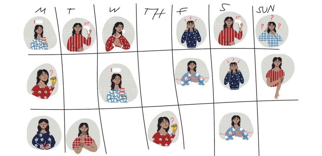
Long COVID patients with fatigue can be tempted to try and push through, to keep speeding through life as they had before their diagnosis. But that doesn’t seem to work for people with chronic fatigue, and “for some long COVID patients, it can actually make things worse,” she’s found.

Azola has similar advice for Hankins. About a half hour into the appointment, Azola slides away from the computer desk, and turns toward her patient. “This is the part where people want to punch me in the face,” she tells Hankins, pushing her glasses up onto her head. “We don’t have a magic wand that makes [you] feel better.”

Instead, Hankins will need to check her body’s battery every day, conserve energy where she can, and build in opportunities to recover. Little tricks, like sitting in a chair while showering or prepping food, can help patients save enough juice to make it through the day. Azola hopes to get Hankins off the “corona coaster,” where patients can feel relatively good one day, and the next day, crash. Having energy levels constantly crater can erode a patient’s ability to live their lives, she says.
For the next 20 minutes, doctor and patient talk about how Hankins’ life has changed and what her next steps will be. In a week, she’ll meet with a neuropsychologist who will help her cope with her new reality; Azola also refers Hankins to a pain specialist.

The two women have spent about an hour together – a near-eternity for a medical appointment. For Azola, it’s time well spent. “The most important thing is to listen to patients and keep an open mind,” she says.

When I speak with Hankins nearly three weeks later, she’s still feeling hopeful. She’s met with the neuropsychologist, and will continue to receive follow-up care. For Hankins, a care plan that factors in all of her conditions, including long COVID, may one day let her feel like herself again.

For now, she’s hoping that sharing her story will help others struggling with the illness. When she tells people she has long COVID, she says, “some of them don’t even think it’s real.”

The pristine Winchcombe meteorite suggests that Earth’s water came from asteroids

Late in the evening of February 28, 2021, a coal-dark space rock about the size of a soccer ball fell through the sky over northern England. The rock blazed in a dazzling, eight-second-long streak of light, split into fragments and sped toward the Earth. The largest piece went splat in the driveway of Rob and Cathryn Wilcock in the small, historic town of Winchcombe.

An analysis of those fragments now shows that the meteorite came from the outer solar system, and contains water that is chemically similar to Earth’s, scientists report November 16 in Science Advances. How Earth got its water remains one of science’s enduring mysteries. The new results support the idea that asteroids brought water to the young planet (SN: 5/6/15).

The Wilcocks were not the only ones who found pieces of the rock that fell that night. But they were the first. Bits of the Winchcombe meteorite were collected within 12 hours after they hit the ground, meaning they are relatively uncontaminated with earthly stuff, says planetary scientist Ashley King of London’s Natural History Museum.
Other meteorites have been recovered after being tracked from space to the ground, but never so quickly (SN: 12/20/12).

“It’s as pristine as we’re going to get from a meteorite,” King says. “Other than it landing in the museum on my desk, or other than sending a spacecraft up there, we can’t really get them any quicker or more pristine.”

After collecting about 530 grams of meteorite from Winchcombe and other sites, including a sheep field in Scotland, King and colleagues threw a kitchen sink of lab techniques at the samples. The researchers polished the material, heated it and bombarded it with electrons, X-rays and lasers to figure out what elements and minerals it contained.

The team also analyzed video of the fireball from the UK Fireball Alliance, a collaboration of 16 meteor-watching cameras around the world, plus many more videos from doorbell and dashboard cameras. The films helped to determine the meteorite’s trajectory and where it originated.

The meteorite is a type of rare, carbon-rich rock called a carbonaceous chondrite, the team found. It came from an asteroid near the orbit of Jupiter, and got its start toward Earth around 300,000 years ago, a relatively short time for a trip through space, the researchers calculate.

Chemical analyses also revealed that the meteorite is about 11 percent water by weight, with the water locked in hydrated minerals. Some of the hydrogen in that water is actually deuterium, a heavy form of hydrogen, and the ratio of hydrogen to deuterium in the meteorite is similar to that of the Earth’s atmosphere. “It’s a good indication that water [on Earth] was coming from water-rich asteroids,” King says.

Researchers also found amino acids and other organic material in the meteorite pieces. “These are the building blocks for things like DNA,” King says. The pieces “don’t contain life, but they have the starting point for life locked up in them.” Further studies can help determine how those molecules formed in the asteroid that the meteorite came from, and how similar organic material could have been delivered to the early Earth.

“It’s always exciting to have access to material that can provide a new window into an early time and place in our solar system,” says planetary scientist Meenakshi Wadhwa of Arizona State University in Tempe, who was not involved in the study.

She hopes future studies will compare the samples of the Winchcombe meteorite to samples of asteroids Ryugu and Bennu, which were collected by spacecraft and sent back to Earth (SN: 1/15/19). Those asteroids are both closer to Earth than the main asteroid belt, where the Winchcombe meteorite came from. Comparing and contrasting all three samples will build a more complete picture of the early solar system’s makeup, and how it evolved into what we see today.

Ancient bacteria could persist beneath Mars’ surface

Radiation-tolerant microbes might be able to live beneath Mars’ surface for hundreds of millions of years and may yet persist today, thanks in part — counterintuitively — to the Red Planet’s frigid, arid conditions.

In addition to being cold and dry, the Martian surface is constantly bombarded by cosmic rays, charged particles and other radiation from space. Previous studies have shown that desiccation vastly extends a microbe’s potential for surviving by limiting the production of highly reactive oxygen-bearing chemicals that can damage proteins and DNA, among other vital molecules within its tissues. To see how long microbes might survive such an onslaught on Mars, researchers desiccated five species of bacteria and one type of yeast, stored them at −80° Celsius and then irradiated them.

Some of the microbes might remain viable for only a few tens of thousands of years, experiments showed. But one species — Deinococcus radiodurans, a particularly radiation-hardy greebly that some scientists have nicknamed “Conan the bacterium” — might survive for as long as 280 million years if protected from radiation at soil depths of 10 meters or more, physical chemist Brian Hoffman and colleagues report online October 25 in Astrobiology.

D. radiodurans resists radiation damage by having multiple copies of chromosomes and other genetic material in each cell, as well as high levels of manganese-bearing antioxidants that help remove DNA-damaging chemicals (SN: 9/3/10). If similar microbes evolved on Mars, they too could persist for lengthy intervals, even possibly until now — which is “improbable but not impossible,” says Hoffman, of Northwestern University in Evanston, Ill.

Even if microbes that evolved on Mars ultimately succumbed to the harsh conditions, remnants of their proteins or other macromolecules may remain — offering hope that future missions, if equipped with the proper equipment, might be able to detect those signs of former life.

A major malaria outbreak in Ethiopia came from an invasive Asian mosquito

In early 2022, malaria cases in the Ethiopian city of Dire Dawa surged, with more than 2,400 people sickened. The spike in infections was the work of an invasive mosquito species that’s spreading across Africa, scientists report.

The finding, presented November 1 in Seattle at the annual meeting of the American Society of Tropical Medicine and Hygiene, provides evidence that the invasive vector can drive malarial outbreaks. Worryingly, the species can thrive in urban environments, bringing the threat of malaria to potentially many millions more people across the continent.

Anopheles stephensi is a mosquito native to India and the Persian Gulf, where it is a major vector for the Plasmodium parasites that cause malaria in people (SN: 10/26/20). In Africa, the primary malaria vector is Anopheles gambiae. A. stephensi was first reported on the African continent in Djibouti in 2012. Since then, the species has turned up in other African countries such as Ethiopia, Somalia and Nigeria.

It wasn’t clear what kind of malarial burden the invasive mosquito could bring to Africa, says Fitsum Girma Tadesse, a molecular biologist at the Armauer Hansen Research Institute in Addis Ababa, Ethiopia. In the eight years after the mosquito’s arrival in Djibouti, the country reported a 40-fold increase in yearly malaria cases, Tadesse says. But no one had directly linked A. stephensi to the increase.

So when malaria cases suddenly rose in Dire Dawa — from 27 cases to 260 in just three weeks in early 2022 — Tadesse and his team jumped in to investigate.

The researchers tracked 80 patients in the city who had sought care for malaria at a local or university clinic, as well as 210 patients who had sought treatment for other reasons, and they screened the patients’ household members for malaria. The team also scanned the patients’ neighborhoods for the presence of mosquito adults and larvae within a 100-meter radius of households, or in the cases of students that visited a clinic, dormitories.

The team found that the malaria patients primarily lived near water sources used by the invasive mosquito, A. stephensi. Households and dorms close to aquatic habitats harboring A. stephensi larvae were 3.4 times as likely as those not close to such water sources to have a family or dorm member test positive for malaria. And most of the adult mosquitoes the team caught — 97 percent — were of the invasive species, the only mosquito species that the researchers found carrying Plasmodium parasites.
A. stephensi “prefers to breed in water storage containers that are typically common in rapidly expanding urban settings,” Tadesse says. The native mosquito species, A. gambiae, tends to use natural sources of water like small pools, which are more common in rural regions, he adds. The concern, then, is that with the expansion of A. stephensi alongside urbanization in Africa, the mosquito could exploit many new sources of water stores.

“This expands the malaria problem from a predominantly rural problem to an urban problem,” says Teun Bousema, an epidemiologist at Radboud University in Nijmegen, the Netherlands.

A 2020 study from another research group estimated that if the invasive mosquito were to spread widely on the continent, an additional 126 million people in cities could be at risk of contracting malaria.

“The spread of Anopheles stephensi is concerning because this species has a number of characteristics that make it difficult to control,” says Tanya Russell, a medical entomologist at James Cook University in Townsville, Australia, who was not involved in the study. Not only can the insects lay their eggs in nearly any available water source, but also the eggs can survive being dry for long periods of time. “This is very uncharacteristic for malaria vectors.”

Insecticide-treated bed nets and spraying a residual insecticide indoors are the primary vector control approaches for malaria-carrying mosquitoes, Russell says. But since A. stephensi also bites outdoors, the mosquito’s spread may blunt the efficacy of these tools.

The key next steps, Tadesse says, are interventions to reduce transmission of the deadly parasites, including targeting the mosquito’s larval phase with chemicals and encouraging communities to cover and secure water containers to prevent mosquitoes from laying eggs in them.

“The window of opportunity to do something about this species is closing,” Bousema says. “So, I really think this calls for very urgent action.”

Clumps of human nerve cells thrived in rat brains

To coax human nerve cells in a laboratory to thrive, there are three magic words: location, location, location.

Many experiments grow human nerve cells in lab dishes. But a new study enlists some real estate that’s a bit more unconventional: the brain of a rat. Implanted clusters of human neurons grow bigger and more complex than their cohorts grown in dishes, researchers report online October 12 in Nature.

Not only that, but the human cells also appear functional, albeit in very limited ways. The implanted human cells can both receive signals from rat cells and influence the rats’ behavior, connections that “demonstrate more substantial integration of the transplanted neurons,” says Arnold Kriegstein, a developmental neuroscientist at the University of California, San Francisco, who wasn’t involved in the study. “This is a significant advance.”

Over the last decade, scientists have been building increasingly complex brain organoids, 3-D clusters of cells derived from stem cells that grow and mimic the human brain (SN: 2/20/18). These organoids don’t re-create the full complexity of human neurons that develop in an actual brain. But they can be windows into an otherwise inscrutable process — human brain development, and how it can go awry (SN: 9/3/21). “Even if they’re not entirely perfect, [these models] are surrogates for human cells in a way that animal cells are not,” Kriegstein says. “And that’s really exciting.”

To push these cells closer to their full potential, Sergiu Pasca, a neuroscientist at the Stanford School of Medicine, and colleagues surgically implanted human cerebral organoids into the brains of newborn rat pups. Along with their hosts, the human organoids began to grow. Three months later, the organoids were about nine times their starting volume, ultimately making up about a third of one side of the rat’s cortex, the outer layer of the brain. “It pushes the rat cells aside,” Pasca says. “It grows as a unit.”

These human cells flourished because rats’ brains offer perks that lab dishes can’t, such as blood supply, a precise mix of nutrients and stimulation from nearby cells. This environmental support coaxed individual human neurons to grow bigger — six times as large by one measure — than the same sort of cells grown in dishes. Cells grown in the rat brains were also more complex, with more elaborate branching patterns and more cell connections called synapses.
The cells looked more mature, but Pasca and his colleagues wanted to know if the neurons would behave that way, too. Tests of electrical properties showed that implanted neurons behaved more similarly to cells that develop in human brains than cells grown in dishes.

Over months of growth, these human neurons made connections with their rat host cells. The human organoids were implanted in the somatosensory cortex, a part of the rat brain that handles whisker input. When researchers puffed air at the whiskers, some of the human cells responded.

What’s more, the human cells could influence the behavior of the rat. In further experiments, the researchers genetically tweaked the organoids to respond to blue light. Prompted by a flash of light, the neurons fired signals, and researchers rewarded the rats with water. Soon, the rats learned to move to the water spout when their human organoid cells sent signals.

In behavioral tests, rats with human implants didn’t show signs of higher intelligence or memory; in fact, researchers were more concerned with deficits. The human organoids were nudging out their hosts’ brains, after all. “Will there be memory deficits? Will there be motor deficits? Will there be seizures?” Pasca asked. But after extensive tests, including behavior tests, EEGs and MRIs, “we could not find differences,” Pasca says.

Other experiments included nerve cells from people with a genetic disorder called Timothy syndrome, a severe developmental disorder that affects brain growth. Growing organoids created with these patients’ cells in rats’ brains might reveal differences that other techniques would not, the researchers reasoned. Sure enough, neurons in these organoids had less complex message-receiving dendrites than those from organoids derived from people without the syndrome.

Organoids made from patient-specific cells could one day even serve as test subjects for treatments, Pasca says. “Challenging disorders will require bold approaches,” he says. “We will need to build human models that recapitulate more aspects of the human brain to study these uniquely human conditions.”

Dinosaur ‘mummies’ may not be rare flukes after all

It might be easier for dinosaurs to “mummify” than scientists thought.

Unhealed bite marks on fossilized dinosaur skin suggest that the animal’s carcass was scavenged before being covered in sediment, researchers report October 12 in PLOS ONE. The finding challenges the traditional view that burial very soon after death is required for dinosaur “mummies” to naturally form.

The new research centers on Dakota, an Edmontosaurus fossil unearthed in North Dakota in 1999. About 67 million years ago, Dakota was a roughly 12-meter-long, duck-billed dinosaur that ate plants. Today, Dakota’s fossilized limbs and tail still contain large areas of well-preserved, fossilized scaly skin, a striking example of dinosaur “mummification.”

The creature isn’t a true mummy because its skin has turned into rock, rather than being preserved as actual skin. Researchers have come to refer to such fossils with exquisitely preserved skin and other soft tissues as mummies.

In 2018, paleontologist Clint Boyd of the North Dakota Geological Survey in Bismarck and colleagues began a new phase of cleaning up and examining the dinosaur fossil. The team had found what looked like tears in the tail skin and puncture holes on the animal’s right front foot. To investigate what may have caused the skin marks, the researchers teamed up with Stephanie Drumheller, a paleontologist at the University of Tennessee in Knoxville, to remove extra rocky material around the marks.

The holes in the skin — particularly those on the front limb — are a close match for bite wounds from prehistoric relatives of modern-day crocodiles, the researchers say. “This is the first time that’s been seen in dinosaurian soft tissues,” Drumheller says.

Because the marks on the tail are larger than those on the front limb, the team thinks that at least two carnivores munched on the Edmontosaurus carcass, probably as scavengers because the wounds didn’t heal. But scavenging doesn’t fit into the traditional view of mummification.

“This assumption of rapid burial has been baked into the explanation for mummies for a while,” Drumheller says. That clearly wasn’t the case for Dakota. If scavengers had enough time to snack on its body, then the deceased dino had been out in the open for a while.

Observing Dakota’s deflated skin envelope, shrink-wrapped to the underlying bone with no muscle or other organs, Drumheller had an unexpected “eureka moment,” she says. “I had seen something like this before. It just wasn’t in the paleontological literature. It was in the forensics literature.”

When some smaller modern scavengers like raccoons feed on the internal organs of a larger carcass, the scavengers rip open the carcass’s body. The forensics research showed that this hole gives any gasses and fluids from further decomposition an escape route, allowing the remaining skin to dry out. Burial could happen afterward.

The researchers “make a very good point,” says Raymond Rogers, a researcher at Macalester College in Saint Paul, Minn., who studies how organisms decay and fossilize and wasn’t involved in the research. “It would be very unlikely for a carcass to achieve advanced stages of desiccation and also experience rapid burial. These two generally presumed prerequisites for mummification seem to be somewhat incompatible.”

Fossilization of soft tissues — like skin or brains or fleshy head combs — is uncommon, but not unheard of (SN: 8/20/21; SN: 12/12/13). “If [soft tissue] requires some spectacular confluence of weird events to get it fossilized at all, it’s far more common than then you would expect if that was the case,” Drumheller says. Perhaps, then, mummies originating from common carcass fates could explain this.

But while dry, “jerkylike” skin could survive long enough to be buried, the conditions involved aren’t necessarily common, says Evan Thomas Saitta, a paleontologist at the University of Chicago who was not involved with the study.

“I still suspect that this actual process is a very precise sequence of events, where if you get the timing wrong, you end up without a mummy dinosaur,” he says.

Understanding that sequence of events, and just how common it is, requires figuring out how fossilization proceeds after a mummy’s burial. This is an area of research that Boyd says he’s interested in looking into next.

“Is it just the same fossilization process as for the bones?” he asks. “Or do we also need a different set of geochemical conditions to then fossilize the skin?”

Drone photos reveal an early Mesopotamian city made of marsh islands

A ground-penetrating eye in the sky has helped to rehydrate an ancient southern Mesopotamian city, tagging it as what amounted to a Venice of the Fertile Crescent. Identifying the watery nature of this early metropolis has important implications for how urban life flourished nearly 5,000 years ago between the Tigris and Euphrates rivers, where modern-day Iraq lies.

Remote-sensing data, mostly gathered by a specially equipped drone, indicate that a vast urban settlement called Lagash largely consisted of four marsh islands connected by waterways, says anthropological archaeologist Emily Hammer of the University of Pennsylvania. These findings add crucial details to an emerging view that southern Mesopotamian cities did not, as traditionally thought, expand outward from temple and administrative districts into irrigated farmlands that were encircled by a single city wall, Hammer reports in the December Journal of Anthropological Archaeology.

“There could have been multiple evolving ways for Lagash to be a city of marsh islands as human occupation and environmental change reshaped the landscape,” Hammer says.

Because Lagash had no geographical or ritual center, each city sector developed distinctive economic practices on an individual marsh island, much like the later Italian city of Venice, she suspects. For instance, waterways or canals crisscrossed one marsh island, where fishing and collection of reeds for construction may have predominated.

Two other Lagash marsh islands display evidence of having been bordered by gated walls that enclosed carefully laid out city streets and areas with large kilns, suggesting these sectors were built in stages and may have been the first to be settled. Crop growing and activities such as pottery making may have occurred there.

Drone photographs of what were probably harbors on each marsh island suggest that boat travel connected city sectors. Remains of what may have been footbridges appear in and adjacent to waterways between marsh islands, a possibility that further excavations can explore.

Lagash, which formed the core of one of the world’s earliest states, was founded between about 4,900 and 4,600 years ago. Residents abandoned the site, now known as Tell al-Hiba, around 3,600 years ago, past digs show. It was first excavated more than 40 years ago.
Previous analyses of the timing of ancient wetlands expansions in southern Iraq conducted by anthropological archaeologist Jennifer Pournelle of the University of South Carolina in Columbia indicated that Lagash and other southern Mesopotamian cities were built on raised mounds in marshes. Based on satellite images, archaeologist Elizabeth Stone of Stony Brook University in New York has proposed that Lagash consisted of around 33 marsh islands, many of them quite small.

Drone photos provided a more detailed look at Lagash’s buried structures than possible with satellite images, Hammer says. Guided by initial remote-sensing data gathered from ground level, a drone spent six weeks in 2019 taking high-resolution photographs of much of the site’s surface. Soil moisture and salt absorption from recent heavy rains helped the drone’s technology detect remnants of buildings, walls, streets, waterways and other city features buried near ground level.

Drone data enabled Hammer to narrow down densely inhabited parts of the ancient city to three islands, she says. A possibility exists that those islands were part of delta channels extending toward the Persian Gulf. A smaller, fourth island was dominated by a large temple.

Hammer’s drone probe of Lagash “confirms the idea of settled islands interconnected by watercourses,” says University of Chicago archaeologist Augusta McMahon, one of three co–field directors of ongoing excavations at the site.

Drone evidence of contrasting neighborhoods on different marsh islands, some looking planned and others more haphazardly arranged, reflect waves of immigration into Lagash between around 4,600 and 4,350 years ago, McMahon suggests. Excavated material indicates that new arrivals included residents of nearby and distant villages, mobile herders looking to settle down and slave laborers captured from neighboring city-states.

Dense clusters of residences and other buildings across much of Lagash suggest that tens of thousands of people lived there during its heyday, Hammer says. At that time, the city covered an estimated 4 to 6 square kilometers, nearly the area of Chicago.

It’s unclear whether northern Mesopotamian cities from around 6,000 years ago, which were not located in marshes, contained separate city sectors (SN: 2/5/08). But Lagash and other southern Mesopotamian cities likely exploited water transport and trade among closely spaced settlements, enabling unprecedented growth, says archaeologist Guillermo Algaze of the University of California, San Diego.

Lagash stands out as an early southern Mesopotamian city frozen in time, Hammer says. Nearby cities continued to be inhabited for a thousand years or more after Lagash’s abandonment, when the region had become less watery and sectors of longer-lasting cities had expanded and merged. At Lagash, “we have a rare opportunity to see what other ancient cities in the region looked like earlier in time,” Hammer says.

How fungi make potent toxins that can contaminate food

Food contaminated with fungi can be an inconvenience at best and life-threatening at worst. But new research shows that removing just one protein can leave some fungal toxins high and dry, and that’s potentially good news for food safety.

Some fungi produce toxic chemicals called mycotoxins that not only spoil food such as grains but can also make us sick. Aflatoxins, one of the more dangerous types of mycotoxins, can cause liver cancer and other health problems in people.

“It is a silent enemy,” says fungal researcher Özgür Bayram of Maynooth University in Ireland, because most people don’t notice when food like corn or wheat is spoiled.

For years, researchers have known that some fungi produce these toxins, but didn’t know all the details. Now, Bayram and colleagues have identified a group of proteins responsible for turning on the production of mycotoxins. Genetically engineering the fungus Aspergillus nidulans to remove even just one of the proteins prevents the toxins from being made, the researchers report in the Sept. 23 issue of Nucleic Acids Research.

“There is a long string of genes that is involved with the production of proteins that, in a cascading effect, will result in the production of different mycotoxins,” says Felicia Wu, a food safety expert at Michigan State University in East Lansing who was not involved in the research.

The newly identified proteins act like a key starting a car, Bayram says. The researchers wanted to figure out how to remove the key and prevent the starting signal from going through, meaning that no toxins would be made in the first place.

Bayram and his team identified the proteins in A. nidulans, revealing that four proteins come together to make the key. The researchers genetically engineered the fungus to delete each protein in turn. When any of the four proteins are missing, the key does not start mycotoxin ignition, the team found.

In another study that has yet to be published, deactivating the same group of proteins in the closely related fungus A. flavus, which can make aflatoxins, prevents the production of those toxins, Bayram says. “So this is a big success because we see, at least in two fungi, the same [protein] complex does the same job.”

The new work “is building upon a body of research that’s been done over decades” to prevent fungal contamination of food, Wu says. A range of methods are already used to control such contamination. For instance, because not all A. flavus strains produce aflatoxins, one method to prevent contamination is to sprinkle nontoxic strains onto fields of corn and peanuts, Wu explains. Those fungi multiply and can help prevent other toxic strains from gaining a foothold.

This research is one of several ways that researchers are using genetic engineering to try to combat these toxins in food (SN: 3/10/17). One future application of the new research could be to genetically tweak a toxin-making fungus and then possibly use it on crops and elsewhere. “We can basically prevent aflatoxin contamination in food, for example, in the field, even in the warehouses, where a lot of contamination takes place,” Bayram says.

Fungi and fungi-like organisms known as water molds are estimated to ruin a third of the world’s food crops each year. If that contamination could be prevented, Bayram estimates the saved food would be enough to feed 800 million people in 2022.

The new research is a good start, Wu says, but it will still be a “challenge to try to understand how this can be operationalized for agricultural purposes.” It’s unclear how scalable the technique is, she says, and getting U.S. regulatory agencies to approve the use of a genetically modified fungus on key food crops might be difficult.

Sharks face rising odds of extinction even as other big fish populations recover

After decades of population declines, the future is looking brighter for several tuna and billfish species, such as southern bluefin tuna, black marlins and swordfish, thanks to years of successful fisheries management and conservation actions. But some sharks that live in these fishes’ open water habitats are still in trouble, new research suggests.

These sharks, including oceanic whitetips and porbeagles, are often caught by accident within tuna and billfish fisheries. And a lack of dedicated management of these species has meant their chances of extinction continue to rise, researchers report in the Nov. 11 Science.

The analysis evaluates the extinction risk of 18 species of large ocean fish over nearly seven decades. It provides “a view of the open ocean that we have not had before,” says Colin Simpfendorfer, a marine biologist at James Cook University in Australia who was not involved in this research.

“Most of this information was available for individual species, but the synthesis for all of the species provides a much broader picture of what is happening in this important ecosystem,” he says.

In recent years, major global biodiversity assessments have documented declines in species and ecosystems across the globe, says Maria José Juan-Jordá, a fisheries ecologist at the Spanish Institute of Oceanography in Madrid. But these patterns are poorly understood in the oceans.

To fill this gap, Juan-Jordá and her colleagues looked to the International Union for Conservation of Nature’s Red List, which evaluates changes in a species’s extinction risk. The Red List Index evaluates the risk of extinction of an entire group of species. The team specifically targeted tunas, billfishes and sharks — large predatory fishes that have influential roles in their open ocean ecosystems.

Red List Index assessments occur every four to 10 years. In the new study, the researchers built on the Red List criteria to develop a way of tracking extinction risk continuously over time, rather than just within the IUCN intervals.

Juan-Jordá and her colleagues did this by compiling data on species’ average age at reproductive maturity, changes in population biomass and abundance from fish stock assessments for seven tuna species, like the vulnerable bigeye and endangered southern bluefin; six billfish species, like black marlin and sailfish; and five shark species. The team combined the data to calculate extinction risk trends for these 18 species from 1950 to 2019.

The team found that the extinction risk for tunas and billfishes increased throughout the last half of the 20th century, with the trend reversing for tunas starting in the 1990s and billfishes in the 2010s. These shifts are tied to known reductions in fishing deaths for these species that occurred at the same time.

The results are positive for tunas and billfishes, Simpfendorfer says. But three of the seven tunas and three of the six billfishes that the researchers looked at are still considered near threatened, vulnerable or endangered. “Now is not the time for complacency in managing these species,” Simpfendorfer says.

But shark species are floundering in these very same waters where tuna and billfish are fished, where the sharks are often caught as bycatch.
“While we are increasingly sustainably managing the commercially important, valuable target species of tunas and billfishes,” says Juan-Jordá, “shark populations continue to decline, therefore, the risk of extinction has continued to increase.”

Some solutions going forward, says Juan-Jordá, include catch limits for some species and establishing sustainability goals within tuna and billfish fisheries beyond just the targeted species, addressing the issue of sharks that are incidentally caught. And it’s important to see if measures taken to reduce shark bycatch deaths are actually effective, she says.

“There is a clear need for significant improvement in shark-focused management, and organizations responsible for their management need to act quickly before it is too late,” Simpfendorfer says.

Zapping tiny metal drops with sound creates wires for soft electronics

Zapping liquid metal droplets with ultrasound offers a new way to make wiring for stretchy, bendy electronics.

The technique, described in the Nov. 11 Science, adds a new approach to the toolbox for researchers developing circuitry for medical sensors that attach to the skin, wearable electronics and other applications where rigid circuit electronics are less than ideal (SN: 6/1/18).

The researchers began by drawing on sheets of stretchy plastic with lines of microscopic droplets made of an alloy of gallium and indium. The metal alloy is liquid at temperatures above about 16° Celsius.

Though the liquid metal is electrically conductive, the droplets quickly oxidize. That process covers each of them with a thin insulating layer. The layers carry static charges that push the drops apart, making them useless for connecting the LEDs, microchips and other components in electronic circuitry.

By hitting the microspheres with high-frequency sound waves, the researchers caused the microscopic balls to shed even smaller, nanoscopic balls of liquid metal. The tiny spheres bridge the gaps between the larger ones, and that close contact allows electrons to tunnel through the oxide layers so that the droplets can carry electricity.

When the plastic that the drops are printed on is stretched or bent, the larger balls of metal can deform, while the smaller ones act like rigid particles that shift around to maintain contact.

The researchers demonstrated their conductors by connecting electronics into a stretchy pattern of LEDs displaying the initials of the Dynamic Materials Design Laboratory, where the work was done. The team also built a sensor with the conductors that can monitor blood through a person’s skin (SN: 2/17/18).

Flexible electronics applications aren’t new, says materials scientist Jiheong Kang of the Korea Advanced Institute of Science and Technology in Daejeon, South Korea. But there are advantages of the new approach over other designs, he says, such as those that rely on channels filled with liquid metal that can leak if the circuitry is damaged. Liquid metal in the conductors that Kang and colleagues developed stays trapped in the tiny spheres that are embedded in the plastic and remains in place even if the material is torn.

Wires made of liquid metal have often been the go-to conductors for stretchy electronics, says Carmel Majidi, a researcher in mechanical engineering at Carnegie Mellon University in Pittsburgh who was not involved with the new study. Using ultrasound introduces a “novel approach to achieving that conductivity.” Other groups have managed that feat by heating circuits, exposing them to lasers, squishing them or vibrating the circuits to get droplets to connect to each other, he says.

Majidi isn’t convinced that the ultrasound approach is a game changer for flexible circuits. But he says that it’s high time the subject is appearing in a leading journal like Science. “I’m personally really excited to see the field overall, and this particular type of material architecture, is now gaining this visibility.”